NAME Advanced fabrication and characterization of biomaterials – CIC biomaGUNE

State-of-the-art infrastructure for the fabrication and characterization of nanostructures mainly applied to molecular imaging, regenerative medicine and biomaterials: • 3D bioprinting • Colloidal nanofabrication of nanoparticles and surfaces • Deposition of biocompatible materials by phase vapor deposition and sputtering • Elemental surface analysis by XPS and UPS • Characterization by NMR • Optical, fluorescence and Raman Microscopy • Transmission and Scanning Electron Miscroscopy • Atomic Force Microscopy • Mass analysis by ICP-MS, UPLC-MS-QToF, MALDI-ToF
FIELDS OF APPLICATION
Additive manufacturing
Biomedical consumables
Electromedical devices
In-Vitro diagnostics
Laboratory equipment
Medical Image
MOST OUTSTANDING EQUIPMENT AND COMPONENTS
-
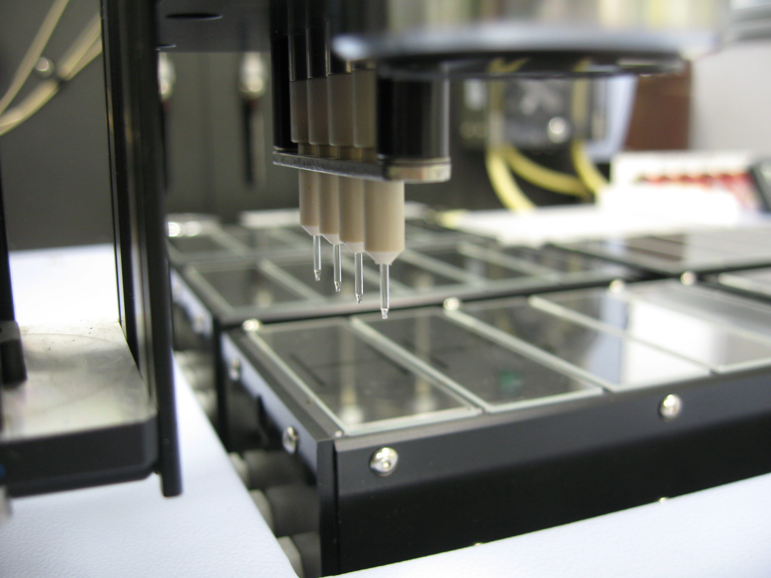
Discovery 3D Evolution BioPrinter
The BioFactory™ instrument is a cutting-edge platform to explore the potential of 3D tissue engineering through bioprinting technology.
Spatial control of cells, bioactives, and extracellular matrix in a three-dimensional cellular construct is an enabling approach to construct designed organomimetic tissues for drug discovery and regenerative medicine.
Laser units for photo polymerization/ biomolecule immobilization.
Modularity: Selection of multiple dispensing technologies.
Biomanufacturing within a sterile laminar flow hood.
Repeatability of micrometer-scale processes.
Fast and easy tissue modelling via Bioprinting Suite. -
Raman Microscope (WITec Alpha 300R)
The Raman microscope is coupled to four different laser excitation sources (488, 532, 633 and 785 nm) and is equipped with two ultrahigh throughput spectrometers with two Peltier-cooled detectors (back-illuminated EMCCD for vis and back-illuminated deep depletion for NIR), four gratings (2x300 grooves/mm for low and 1800 and 1200 grooves/mm for high spectral resolution) optimized for visible and NIR range.
The topographic sensor allows to measure on rough surfaces always "in-focus". The motorized scanner stage has a maximal travel distance of 50 x 50 mm2. -
Transmision Electron Microscope TEM – JEOL JEM-2100F (UHR 2007)
Is a Transmission Electron Microscope equipped with a field-emission gun (FEG) transmission with variable acceleration between 80 and 200 kV, a 4-megapixel CMOS camera (TVIPS TemCam-F216) and two STEM detectors (BF & HAADF) in combination with an EDX detector (Oxford UltimMax updated in 2020), and has the GATAN 626 cryo-transfer system
-
X-ray Photoelectron Spectroscope XPS/UPS – SPECS SAGE HR-100
It is an X-ray Photoelectron Spectroscope equipped with a 100 mm radius PHOIBOS analyser. It allows to perform analysis of elemental composition and determine the energy of the valence levels up to a depth of 5 to 10 nm. It has two non-monochrome x-ray sources (Al and Mg K-) and a UVS 10/35 ultraviolet lighting source (updated in 2020). Surface layers can be removed from substrates by using Argon ion plasma, as well as making a composition profile based on applied time.
-
Zeiss LSM 880 Confocal Multiphoton Fluorescence Microscope.
Zeiss LSM 880 Airyscan and MaiTai DeepSee (updated in 2021). It is equipped with three Ar laser excitation sources (458, 488 and 514 nm), DPSS (561 and 590 nm) and HeNe (633 nm). It is coupled to a MaiTai DeepSee multiphoton laser, with variable excitation range (690 to 1040 nm). It has two PMT photomultiplier type detectors, one standard and one with high sensitivity in the infrared range, plus a GaAsP detector for visible detection. It also has a BIG-2 detector module, consisting of two GaAsP detectors for high sensitivity multiphoton measurements. Allows measurements of correlation spectroscopy and cross-correlation of fluorescence.
SERVICES OFFERED BY THE ASSET
3D Bioprinting
Design and 3D printing of complex composite structures with biological properties. The BioFactory™ is a versatile and cell friendly three-dimensional manufacturing instrument allowing to pattern cells, biomolecules and a range of soft and rigid materials in desirable 3D composite structures, mimicking natural environments.
Atomic Force Microscopy of biomaterials
Determination of surface roughness, grain size or nanoparticle size on surfaces and substrates, nano-mechanical properties of surfaces. Applicable to the study of different substrates such as: films, polymers, cells, proteins, viruses, bacteria, biomolecules, etc.
Colloidal Nanofabrication with plasmonic resonance properties
Development of synthetic strategies to obtain structurally uniform metallic nanoparticles in high yield, with excellent control over their shape and size, and a wide variety of surface functionalization. Different experimental pathways are available for the preparation of a wide variety of hybrid nanomaterials.
Electron Microscope Characterization of biomaterials
Preparation of (cryo)samples and corresponding (cryo)microscopy studies. A dedicated sample preparation room is equipped to perform immersion freezer, high pressure freezer, automatic freezing replacement and an ultramicrotome with an accessory to process samples under cryogenic conditions.
Elemental composition analysis of surfaces by X-ray Photoelectron Spectroscopy XPS
XPS allows to determine the composition (qualitatively and quantitatively) of the most superficial layer of a material. It allows to determine the chemical state of the elements present on the surface of the material. Study of corrosion, asses the stability of the substrates or determine the presence of impurities.
Optical Microscopy Characterization of samples
Characterization of materials by optical and spectroscopical techniques of biomaterials and biological systems in vitro and ex vivo. It allows obtaining transmission, reflection and fluorescence images of biomaterials and biological samples with low or high resolution, both laterally and in depth.
Optical Spectroscopic Characterization of samples
Characterization of the optical properties of compounds and biomolecules in solution, suspension or solid state. Measurement of extinction or transmission coefficients of solutions, dispersions or other partially transparent materials. Multimode measurement of fluorescence, phosphorescence and intensity of absorbance and transmission.
ENTITY MANAGING THE ASSET

Contact person:
Angel Martínez
external.services@cicbiomagune.es